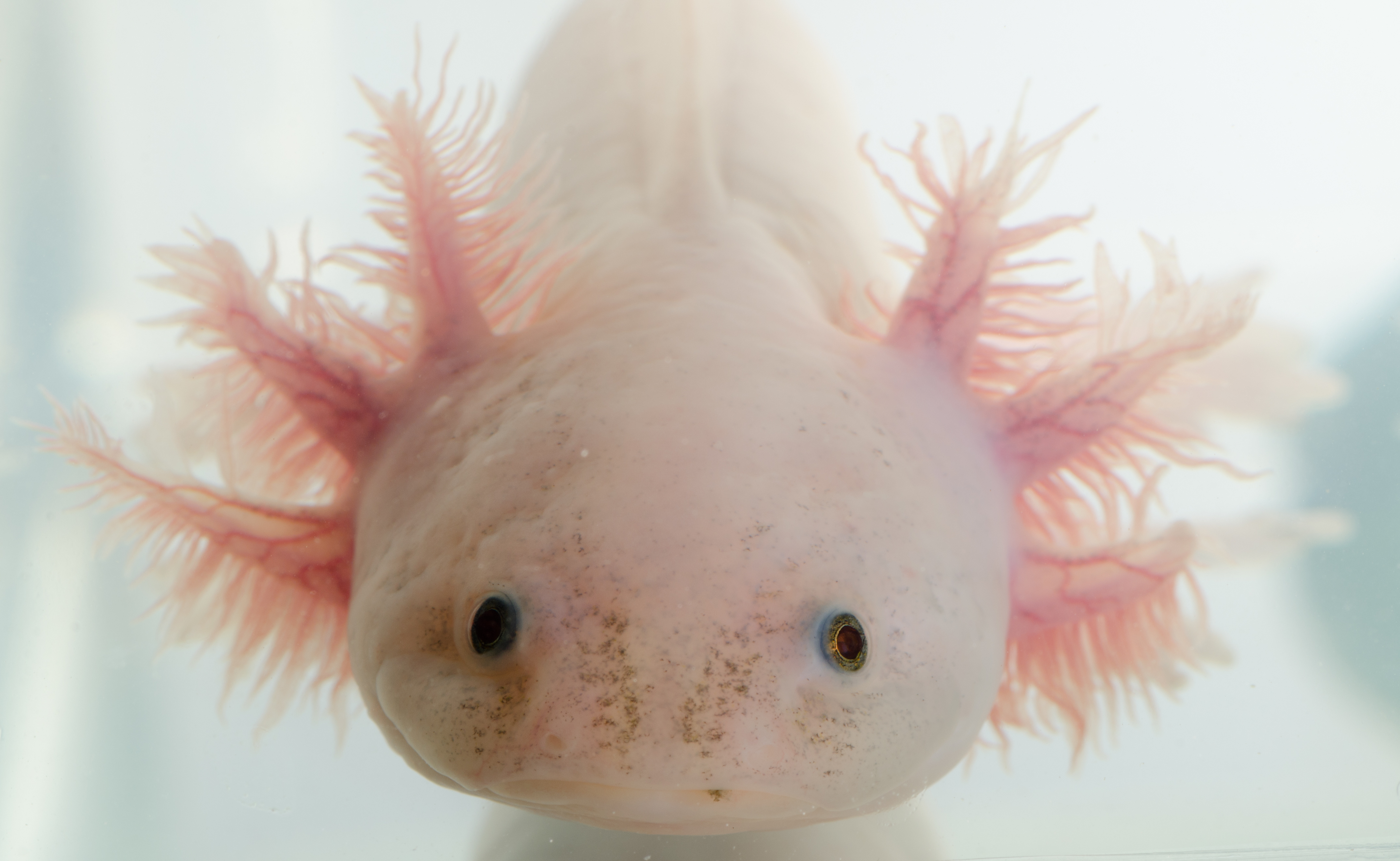
The latest cover story in Science on the BGI-led groundbreaking research into brain development and regeneration in the axolotl salamander has attracted international scientific attention and demonstrates how spatiotemporal technology can be applied to transform mankind’s understanding of the world around us and bring new advancements in disease research.

Described by Dr Elly Tanaka, Biochemist and Senior Scientist at The Research Institute of Molecular Pathology in Vienna, Austria, as “a fantastic new spatial transcriptomic technology from BGI”, Stereo-seq enables scientists to map a panoramic atlas of every cell in an organism according to their individual biomolecular profile, in space and over time, and to do this very efficiently and very quickly.
In the latest research published by Science, this technology was used to create a single-cell resolution spatiotemporal map of salamander brain development over six important developmental periods, showing the molecular characteristics of various neurons and dynamic changes in spatial distribution.
Researchers found that neural stem cell subtypes located in the ventricular zone region in early stage of development are difficult to distinguish, but began to specialize with spatial regional characteristics from the adolescent stage. This discovery suggests different subtypes may undertake different functions during regeneration.
By sampling the brain at seven time points (2, 5, 10, 15, 20, 30 and 60 days) following an injury to the cortical area of the salamander brain, the researchers were able to analyze cell regeneration.
"Using axolotl as a model organism, we have identified key cell types in the process of brain regeneration. This discovery will provide new ideas and guidance for regenerative medicine in the mammalian nervous system," explained Dr. Ying Gu, joint corresponding author of the paper, Deputy Director of BGI-Research.
In the early stage of injury, new neural stem cell subtypes began to appear in the wound area, and partial tissue connections appeared in the injured area on day 15. On days 20 and 30, researchers observed that the wound had been filled with new tissues, but the cell composition was significantly different from the non-injured area. By day 60, the cell types and distribution had returned to the same status as the non-injured area.
“It is no exaggeration that this study has indicated a very important direction for us to investigate the repair of brain injuries in mammals, especially in primates and humans in the future,” says Dr Chengyu Li, Senior Investigator at the Center for Excellence in Brain Science and Intelligence Technology of the Chinese Academy of Sciences. “It has profound meaning for the development of treatments for brain injuries.”
Dr Maria Antonietta Tosches, Assistant Professor in the Department of Biological Sciences at Columbia University, agrees: “This paper really demonstrates how new technologies such as Stereo-seq that has unprecedented spatial and single-cell resolution, can open-up new insights on long-standing biological questions such as brain regeneration.”
Dr An Zeng, Investigator at the Center for Excellence in Molecular Cell Science of the Chinese Academy of Sciences, adds: “Such technology is important for the research on regeneration of other tissues and organs or regeneration models.”
Dr Tosches concludes, “We are really excited about the paper and new technology, and are really looking forward to seeing the new research that it will open up in the scientific community.”
In the past six months BGI's research achievements in spatiotemporal omics and single-cell technology have been published four times in the leading scientific journals "Cell", "Nature" and "Science”.
Watch the video:


