


The 2026 “Chinese+Intelligent Bioengineering” DUT-BGI International Summer School, jointly organized by BGI College and Dalian University of Technology, officially opened on July 27 at the BGI Center in Shenzhen, China.
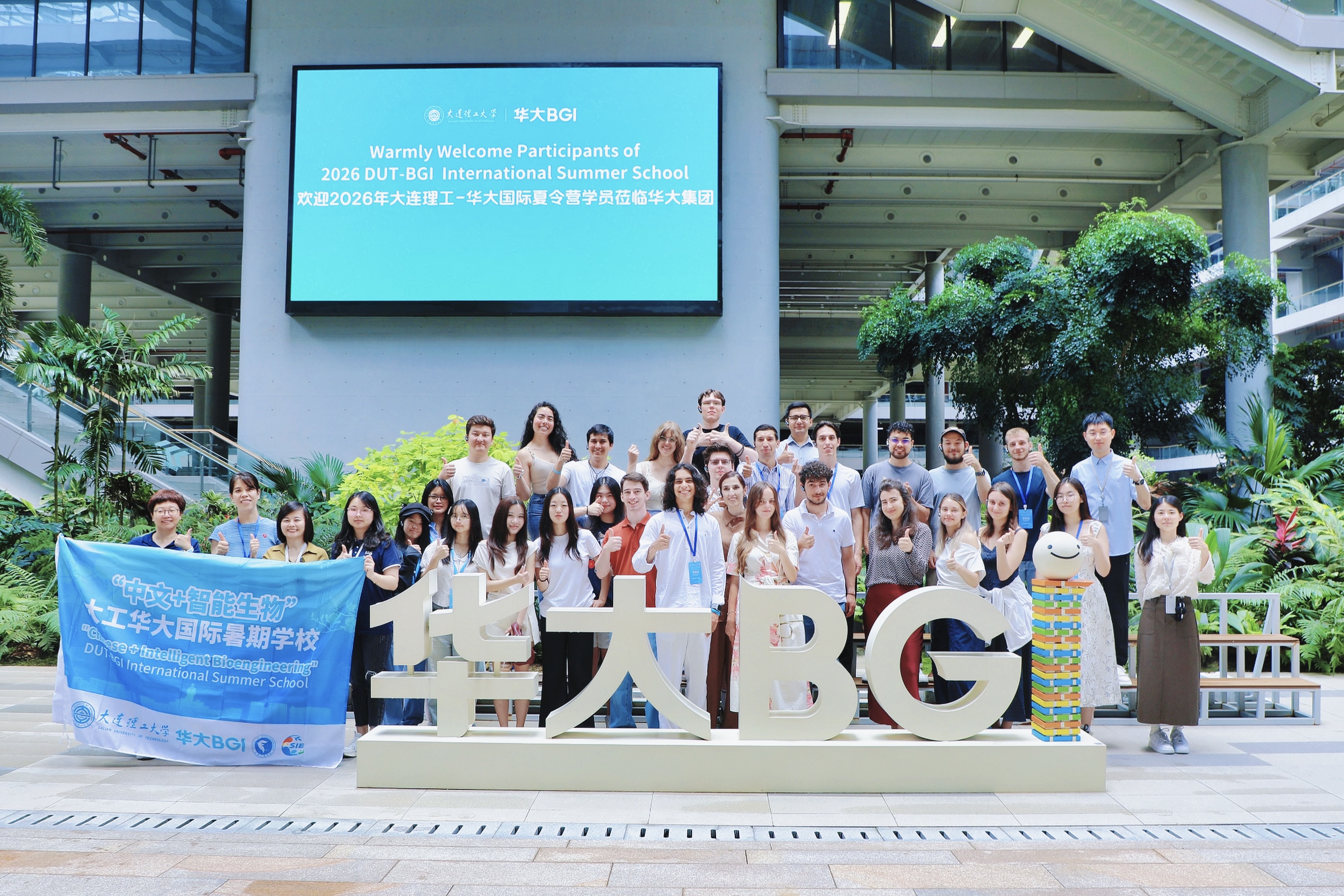 Group photo of the 2026 DUT-BGI International Summer School participants.
This year’s program brings together 30 young students and scholars from 10 countries, including Colombia, Uzbekistan, Serbia, and Indonesia, for a one-week program focused on life sciences, genomics innovation, active health, and international research collaboration.
The summer school is designed to provide participants with an integrated learning experience through lectures, visits to research platforms, laboratory practice, case studies, academic exchanges, and group discussions. The program aims to help young participants better understand the development of life sciences technologies, their practical applications, and opportunities for global cooperation.
On the opening day, participants visited the BGI Center, where they were introduced to BGI Group’s development history, major scientific research achievements in life sciences, global research platform layout, and the 133111i active health management system. The visit provided an overview of BGI’s work in genomics, research platform development, technology application, and industrial transformation.
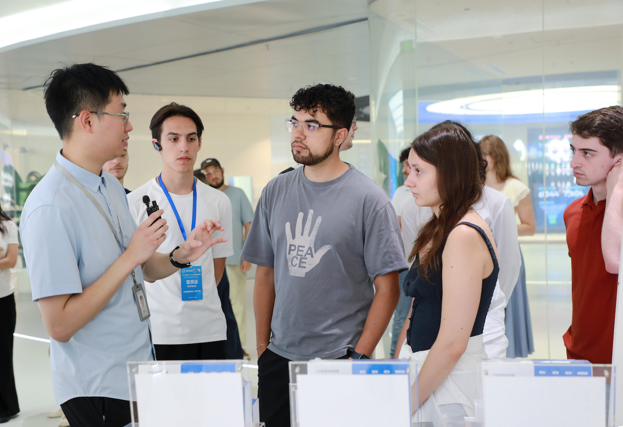 Participants visit BGI Future Intelligent Health Center in the BGI Center.
The opening ceremony also included exchanges among participants from different countries and academic backgrounds. Students and scholars shared their areas of study, research interests, and learning experiences, covering fields such as bioengineering, artificial intelligence, and related interdisciplinary areas.
Over the coming week, participants will take part in courses and activities based on BGI’s research and technology platforms. The curriculum will cover BGI Group’s technologies and products, the development of sequencing technologies, genomics, and large-scale scientific programs. Participants will also discuss real-world global cases and explore how life sciences technologies can support healthcare needs in different countries and regions.
In addition to academic activities, the program includes sessions related to active health and healthy lifestyles, helping participants better understand the connection between scientific research, health management, physical activity, nature, and daily life.
The 2026 DUT-BGI International Summer School serves as a platform for international education, scientific exchange, and collaboration among young researchers. Through the program, BGI Group aims to support global talent development in life sciences and promote dialogue and cooperation among young students and scholars from different countries.