



On May 27, 2026, a research team led by the Obstetrics and Gynecology Hospital of Fudan University and the State Key Laboratory of Genome and Multi-omics Technologies at BGI-Research, in collaboration with Zhejiang University School of Medicine and other institutions, reported in Nature a spatiotemporal transcriptomic atlas of whole human embryos spanning Carnegie stages (CS) 12 to 23 — equivalent to four to eight weeks post-conception. This critical period witnesses rapid organogenesis, and developmental perturbations may lead to lifelong conditions including birth defects and neurodevelopmental disorders.
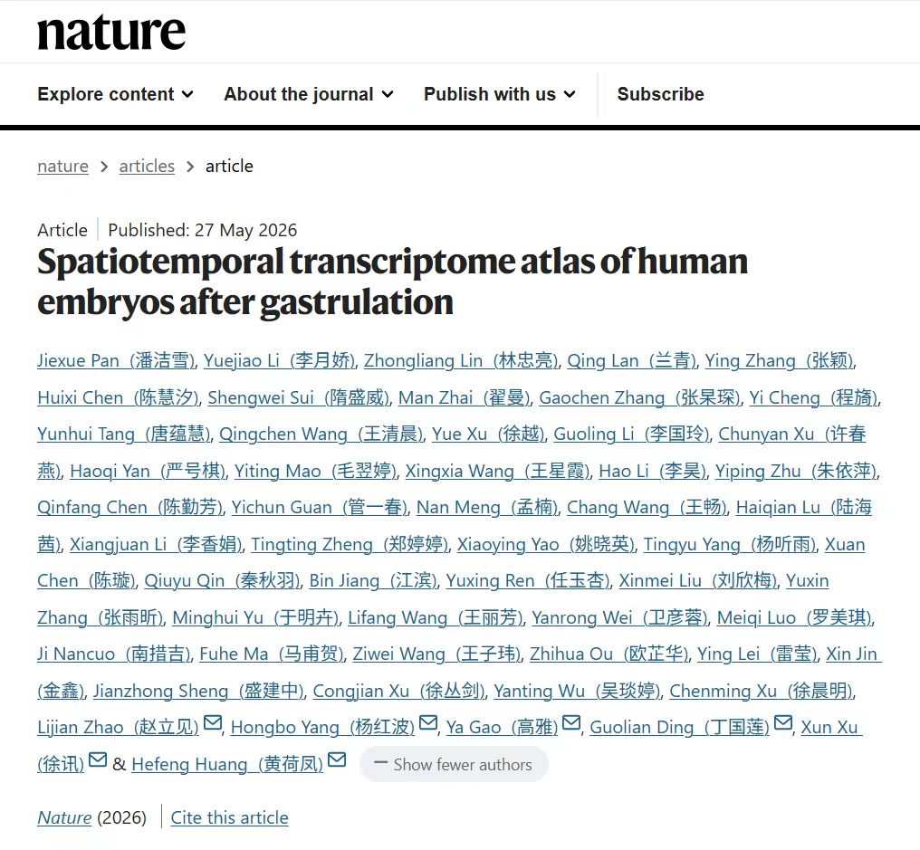 The study titled Spatiotemporal transcriptome atlas of human embryos after gastrulation was published in Nature.
Combining Stereo-seq spatial transcriptomics and single-nucleus RNA sequencing, the team generated data on the DNBSEQ platforms with single-nucleus libraries constructed using DNBelab C4 kits. They profiled 77 sagittal sections from 13 euploid embryos and more than 600,000 single nuclei, mapping gene expression across 50 organs and 198 molecularly defined substructures. This atlas fills a long-standing gap between gastrulation research and fetal organ analysis, providing a spatial and regulatory reference for human organogenesis. The resource is publicly accessible via the HESTA portal (https://db.genomics.cn/hesta).
One key finding from the atlas relates to the regulatory network governing early heart development. From 58 cardiac sections, the researchers annotated 26 substructures and precisely located the sinoatrial node — the heart’s natural pacemaker — at CS17. Beyond well-characterized pacemaker regulators such as SHOX2, two additional genes were found to be highly enriched in the sinoatrial node: KIAA1324L, which had not previously been linked to cardiac function, and RORA, a gene primarily known for regulating circadian rhythms. In situ hybridization validated their expression alongside the canonical pacemaker marker HCN4. Functional assays via CRISPR–Cas9 knockout of their orthologs in zebrafish resulted in reduced pacemaker cell numbers and lowered heart rate, confirming their functional roles in sinoatrial node development.
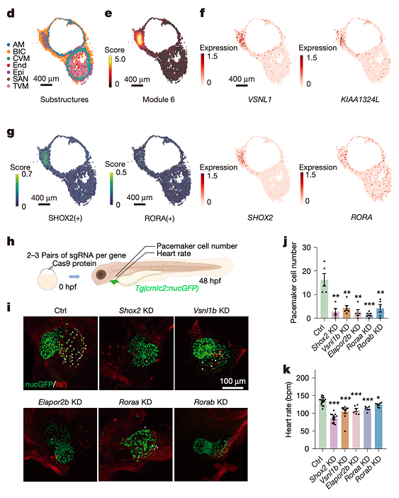
The atlas pinpoints the embryonic sinoatrial node and identifies KIAA1324L and RORA as candidate regulators of cardiac pacemaking, with zebrafish perturbation reducing pacemaker cells and heart rate.
The work also refines the timeline of early neurogenesis in the embryonic brain. Markers for inhibitory neurons were detected in the ganglionic eminence as early as CS12–13, earlier than documented in previous studies, while the specification of excitatory neurons occurred at a later stage. Notably, the study identified HMGA2 as a dynamic regulator in the pallial ventricular zone. In silico gene perturbation analyses indicated that loss of HMGA2 accelerates cell differentiation at the expense of radial glial cell maintenance.
Furthermore, HMGA2 showed a rostral-dominant pattern in humans, in contrast to caudal enrichment in mice, a divergence that may provide insights into species-specific features of cortical development. Because the HMGA2 network includes downstream targets linked to intellectual disability, these findings may help researchers explore how disruptions during early brain development contribute to neurodevelopmental conditions.
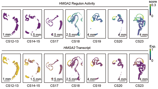
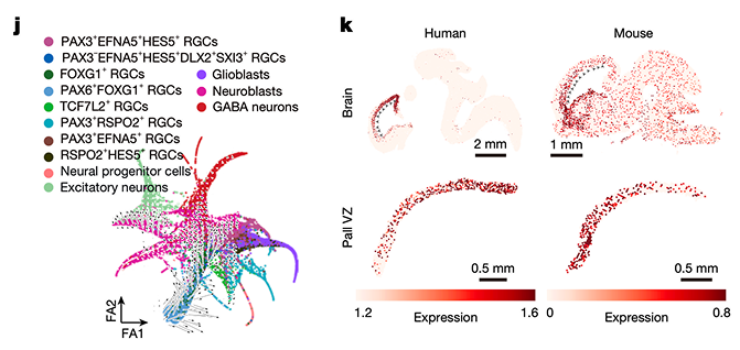
Spatial maps of HMGA2 regulon activity and transcript expression reveal ventricular-zone enrichment after CS17, while perturbation simulation and human–mouse comparison link radial-glial-cell maintenance to species-specific cortical patterning.
The atlas also resolves how organ substructures emerge along developmental trajectories. In the developing eye, pseudotime analysis traced retinal progenitor cells as they branched toward melanocytic and neurogenic lineages, with transcription factors MITF, ISL1, and KLF7 showing coordinated shifts in expression and regulon activity that aligned with distinct cell-fate transitions. These regulatory profiles may help researchers explore the cellular origins of congenital eye malformations.
In the kidney, the team reconstructed nephron progenitor differentiation toward stromal, podocyte, and tubular fates, with regulators such as HNF1B and MAFB displaying distinct enrichment across renal substructures. Together, the eye and kidney analyses illustrate that organogenesis proceeds through spatially localized regulatory relays.
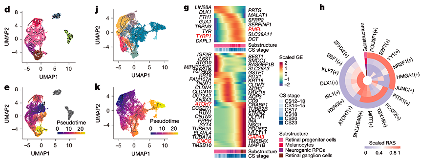
Pseudotime and regulon analyses trace retinal progenitor branching and nephron lineage specification, revealing coordinated regulators of eye and kidney substructure formation.
The atlas further connects development with disease susceptibility. Cytomegalovirus receptor genes such as CD147 and THY1 were detected in the embryonic brain and inner ear as early as CS12–13, while SARS-CoV-2 entry factors ACE2 and TMPRSS2 were largely restricted to gut epithelium at CS23 with rare same-cell co-expression; the authors noted that receptor expression alone cannot fully explain tropism or transmission risk.
Analysis of 1,740 disease-associated genes from the DDG2P database revealed organ-specific enrichment matching known clinical phenotypes. Cross-species comparisons identified human–mouse differences for genes such as ARG1 and CCBE1, underscoring the value of human-specific developmental references.
The team additionally identified 452 genes with allelic imbalanced expression, including known imprinted genes and novel candidates. This expands the atlas beyond average gene activity into a reference for allele-biased and parent-of-origin regulation.
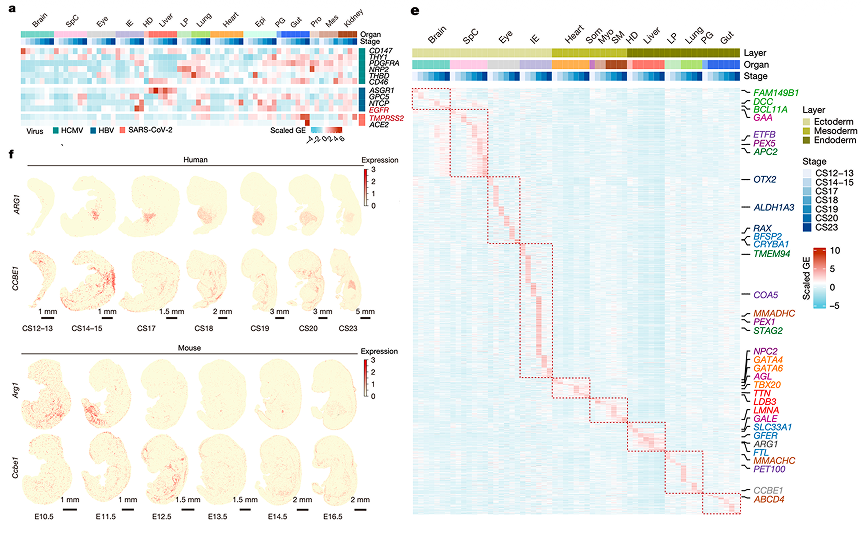
Viral-entry factors, DDG2P developmental-disease genes, and ARG1/CCBE1 human–mouse differences reveal organ- and stage-specific vulnerability patterns during early embryogenesis.
This multi-dimensional atlas integrates molecular, cellular, organ-level, cross-species and allelic regulatory landscapes of human organogenesis into a unified spatial framework. It serves as a foundational reference for research into birth defects, congenital infection, and neurodevelopmental disorders, and as a benchmark for in vitro models such as organoids. It also demonstrates the capacity of Stereo-seq for large-scale, high-resolution spatial mapping of developmental processes.
All raw data and resources are available through HESTA, GEO, GSA-Human, and CNGBdb. All studies involving human embryos and zebrafish were approved by institutional ethics committees, and all human tissue donors provided written informed consent.
This research can be accessed at https://doi.org/10.1038/s41586-026-10545-0.