



In a study recently published in Science China Life Sciences, investigators from State Key Laboratory of Genome and Multi-omics Technologies at BGI-Research and collaborating institutions report the construction of a high-resolution single-cell multi-omics atlas of human immune aging that uncovers extensive, previously hidden changes in RNA transcript architecture and immune receptor diversity. By integrating gene expression, full-length transcript isoform sequencing, and immune receptor profiling from the same individual cells, the study reveals molecular layers of immunosenescence, the progressive decline of immune function with age, that conventional methods have been unable to detect.
 The study “Single-Cell Multi-Omics Dissects Transcript Isoform and Immune Repertoire Dynamics in Human Immunosenescence” was published in Science China Life Sciences.
The research team profiled peripheral blood immune cells from 19 healthy adults (9 aged 30 to 40 and 10 aged 60 to 70) using three complementary single-cell sequencing approaches linked by shared cell barcodes. This strategy yielded approximately 104,000 high-quality single cells resolved into 30 immune cell subtypes, capturing 78,401 transcript isoforms and fully reconstructed T cell and B cell receptor repertoires. The long-read sequencing component alone identified 17,466 novel transcript isoforms absent from the reference annotation, demonstrating that a substantial layer of immune cell biology remains invisible to standard short-read methods.
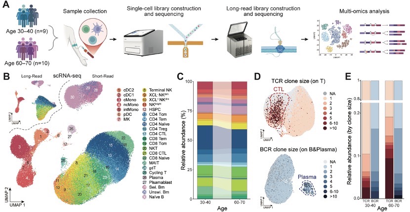 A single-cell multi-omics atlas of approximately 104,000 immune cells resolves 30 subtypes and reveals age-associated changes in cell composition and T cell clonal expansion.
A central finding emerged from the transcript isoform analysis: within CD4+ effector memory T cells, the researchers identified a senescence-prone subpopulation enriched in older individuals. These cells preferentially expressed a truncated version of the HSPA5 gene, which encodes a molecular chaperone critical for handling protein-folding stress during T cell activation. Structural analysis predicted that the truncated transcript is functionally incompetent, lacking essential domains and likely subject to degradation, potentially leaving these aging T cells less equipped to manage the protein-folding stress that accompanies immune activation.
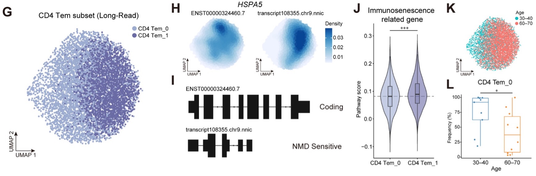 Within CD4+ effector memory T cells, a senescence-prone subpopulation enriched in older adults preferentially expresses a truncated HSPA5 isoform predicted to impair stress management during immune activation.
Beyond individual gene “version switches,” the study uncovered systematic changes in the regulatory tails of RNA messages across aging immune cells. Older individuals expressed transcripts with longer 3’ untranslated regions (3’UTRs), particularly in CD4+ T cells. These extended tails introduce additional docking sites for small regulatory molecules called microRNAs, which can silence gene activity. The result functions as a set of hidden brakes on immune gene expression: longer 3’UTRs on genes such as GADD45B (involved in stress response and DNA damage repair) and ZFP36L2 (an immune gene regulator) introduce additional microRNA binding sites that can suppress their expression, suggesting that this post-transcriptional remodeling may broadly dampen immune cell function during aging.
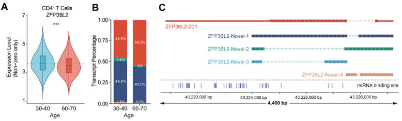 Reduced ZFP36L2 expression in aging CD4+ T cells coincides with shifts toward novel transcript isoforms carrying extended 3’UTRs and additional microRNA binding sites.
The atlas also revealed a striking functional transformation in cytotoxic T lymphocytes (CTLs), the immune system’s specialized cell-killing force. In older individuals, the most expanded CTL clones had shifted away from their classical cell-killing programs toward an inflammatory state driven by NF-κB and type I interferon signaling pathways. Rather than defending the body against threats, these clonally expanded cells appear to have been reprogrammed into drivers of chronic, low-grade inflammation, a hallmark of aging known as inflammaging. T cell receptor diversity narrowed markedly with age in these subsets, with expanded clones exhibiting elongated receptor recognition regions consistent with chronic stimulation by persistent antigens.
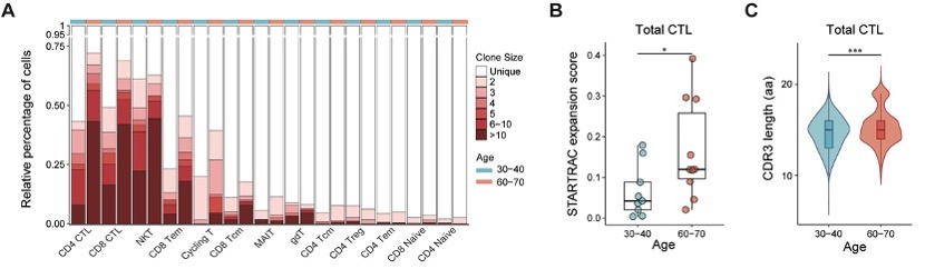 Cytotoxic T lymphocytes in older individuals show pronounced clonal expansion and elongated receptor recognition regions, consistent with chronic antigenic stimulation driving immune repertoire narrowing.
Notably, not all arms of adaptive immunity followed the same trajectory. B cell receptor repertoires and somatic hypermutation, the process by which B cells refine their ability to recognize pathogens, remained largely comparable between the two age groups, suggesting that B cell maturation machinery retains considerable resilience during this window of aging.
Because scCycloneSEQ full-length transcriptome sequencing and DNBelab C series single-cell capture share cell barcodes, the researchers could link each cell’s gene expression, transcript isoform structure, and immune receptor identity in a single integrated framework, directly connecting clonal expansion patterns to the transcript-level changes that accompany them.
“This atlas is an early output of our CIMA-II initiative for immune multi-omics research,” said corresponding author Chuanyu Liu from BGI-Research. “We plan to extend this framework to autoimmune and cardiovascular diseases, and the high-quality dataset will serve as a core foundation for virtual immune cell modeling.”
Extending this multi-omics approach to tissue-resident immune populations, developmental stages, and longitudinal cohorts could further clarify how transcript-level changes unfold over time and across different tissue environments. By establishing that immune aging operates through hidden regulatory layers well beyond gene expression levels, the atlas provides a new foundation for identifying candidate mechanisms that may one day inform strategies to preserve immune function in aging populations.
This study was approved by the relevant institutional review board, and all participants provided written informed consent in accordance with applicable human research ethics regulations.
This research is available at: https://www.sciengine.com/SCLS/doi/10.1007/s11427-025-3398-8