



 The study “Spatiotemporal multiomics uncover tumor ecosystem dynamics during metastatic colonization” was published in Science.
Metastasis, the spread of cancer to distant organs, remains the leading cause of cancer-related death. Yet the earliest steps of metastatic colonization, when cancer cells arrive in a new organ and most are rapidly eliminated, have been exceedingly difficult to study. The surviving cells are rare, their molecular programs shift quickly, and conventional single-cell methods lack the spatial context to capture these fleeting events. To address this, the study team constructed a detailed molecular “time-lapse” of lung colonization by hepatocellular carcinoma (HCC) cells in immunocompetent mice. Lungs were sampled at nine time points, from 15 minutes to 35 days after tumor cell arrival, with seven preceding visible metastatic outgrowth.
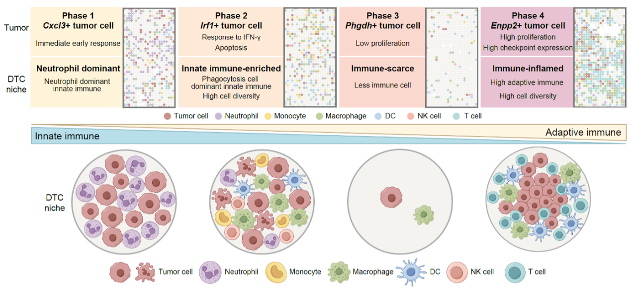 Metastatic colonization proceeds through four phases: early immune response, innate clearance, a quiescent immune-scarce niche marked by Phgdh-high tumor cells, and immune-inflamed outgrowth.
The researchers integrated Stereo-seq spatial transcriptomics, a technology that reads gene activity while preserving each cell’s exact location within the tissue, with single-cell RNA sequencing and chromatin accessibility profiling. Stereo-seq proved essential for detecting the rare surviving tumor cells during the critical micrometastatic window: the platform captured over 2,000 tumor cells at stages when conventional single-cell sequencing detected fewer than 20. The atlas encompassed more than 7.5 million spatial spots (10 × 10 μm2) from 32 lung sections, alongside single-cell profiles from over 180,000 cells and 284,000 nuclei.
Analysis of this atlas revealed that metastatic colonization proceeds through a coordinated sequence of ecological states rather than a simple expansion in tumor cell number. After most disseminated tumor cells (DTCs) were eliminated by neutrophils and natural killer cells within hours of arrival, a small residual population survived by entering a transient, low-proliferative state marked by high expression of the metabolic enzyme PHGDH. Rather than fueling growth, as previously described in primary tumors, PHGDH in these surviving cells drove production of the methyl-donor molecule SAM (S-adenosylmethionine). Elevated SAM, in turn, promoted a repressive chemical tag on DNA-packaging proteins, H3K27me3, which acted as a molecular “mute switch” to silence chemokine genes, the signals that would normally recruit immune cells to attack. The result was an immune-scarce niche in which the surviving cancer cells could persist largely undetected.
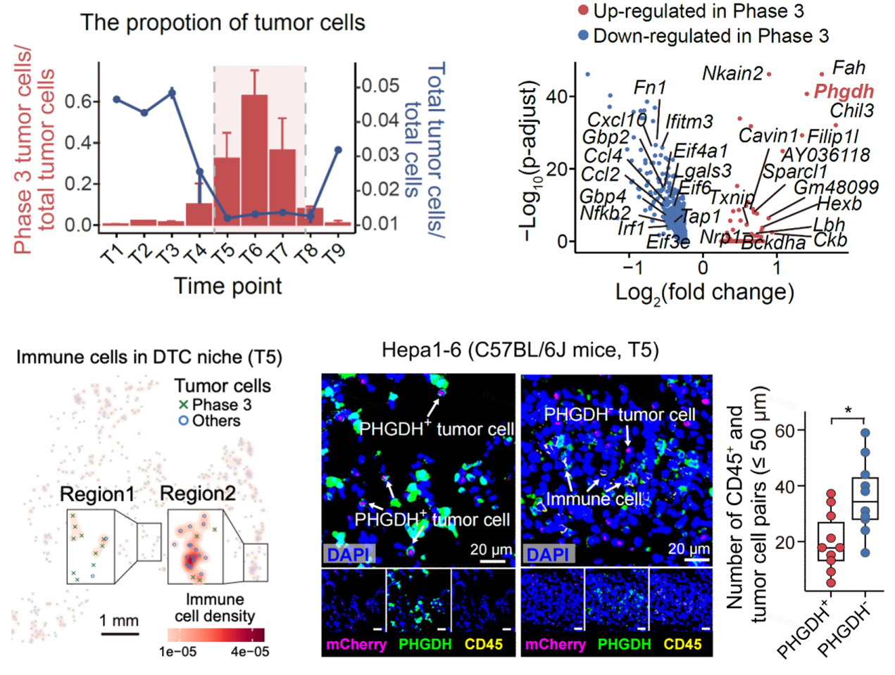 Transient Phgdh-high tumor cells occupy niches with markedly fewer nearby immune cells during the micrometastatic phase, as shown by spatial profiling and multiplex immunofluorescence.
Lineage tracing in the mouse model demonstrated that the majority of cells in later, overt metastases descended from ancestors that had transiently passed through this Phgdh-high state. The study describes these cells as an adaptive phenotype induced by microenvironmental cues rather than a predetermined subpopulation from the primary tumor. Observations in human metastatic specimens supported cross-species relevance: a small subset of Phgdh-high tumor cells was identified in immune-scarce niches across liver, breast, colorectal, and lung cancer metastases, and higher Phgdh-high signature scores were associated with worse clinical outcomes in retrospective cohorts.
The study uncovered a second, sequential mechanism. Before metastatic outgrowth began, Cx3cr1-high interstitial macrophages accumulated in the tumor niche, recruited by tumor-derived CX3CL1. These macrophages reorganized the local environment by attracting immunosuppressive cells, including regulatory T cells and immature neutrophils, and provided growth signals through the IGF1 pathway in ex vivo experiments, collectively creating conditions supporting the transition from quiescent micrometastasis to active expansion.
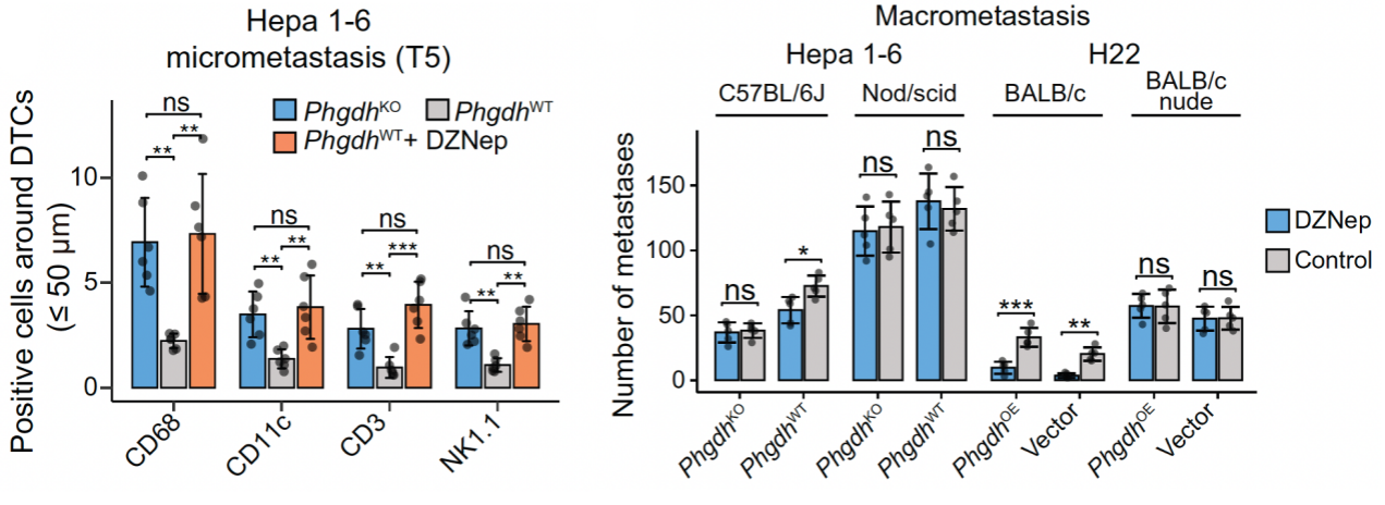 Genetic loss of Phgdh or pharmacological inhibition of SAM-dependent histone methylation increased immune infiltration and reduced metastatic burden in immunocompetent, but not immunodeficient, mice.
In preclinical experiments, disrupting either mechanism suppressed metastatic colonization. Genetic or pharmacological perturbation of the PHGDH-H3K27me3 axis restored chemokine expression and immune cell recruitment, significantly reducing metastatic burden in immunocompetent mice; this effect was absent in immunodeficient mice, supporting an immune-mediated mechanism. Depleting interstitial macrophages during the pre-outgrowth window reduced immunosuppressive cell infiltration and metastatic colonization.
The findings suggest that micrometastases and established metastases have fundamentally different molecular programs, and that some of the decisive vulnerabilities of early colonization disappear once a visible lesion has formed. Extending these approaches to additional cancer types and, ultimately, to clinical settings could open new avenues for intercepting metastatic disease at its most vulnerable stages, before it becomes visible to current diagnostic methods. The full spatiotemporal dataset is publicly available through the Cancer Omics Atlas of Spatiotemporal Topology (COAST) interactive visualization portal, providing a resource for the broader cancer research community.
Ethics approval for this study is obtained. All clinical specimens were collected with informed consent.
This research is available at: https://doi.org/10.1126/science.adz7928