



On August 18, 2026, researchers from State Key Laboratory of Genome and Multi-omics Technologies at BGI-Research, Jilin University, and Jinan University report in Cell a cell-by-cell survey of the aging primate brain showing that the heaviest burden of age-related gene-expression change falls on a white-matter-rich stretch of pons and medulla. The same data show that the pace of change is far from even, and that the oldest animals were not simply the most worn down.
 The study titled “Multimodal brain cell atlas across the adult macaque lifespan” was published in Cell.
The circuits aging hits hardest are far more expanded and specialized in primates than in rodents, and human tissue is variable and scarce at the oldest ages, so the team went to the cynomolgus macaque, profiling close to three million cell nuclei from eight brain regions in 23 females across four age groups covering ages 5 to 31 - with the oldest animals approaching the upper end of this species' typical lifespan in captivity. Nuclei from each sample were split between two assays, one reading which genes a nucleus had switched on, the other which stretches of its DNA lay open, with sequencing carried out on DNBSEQ platforms.
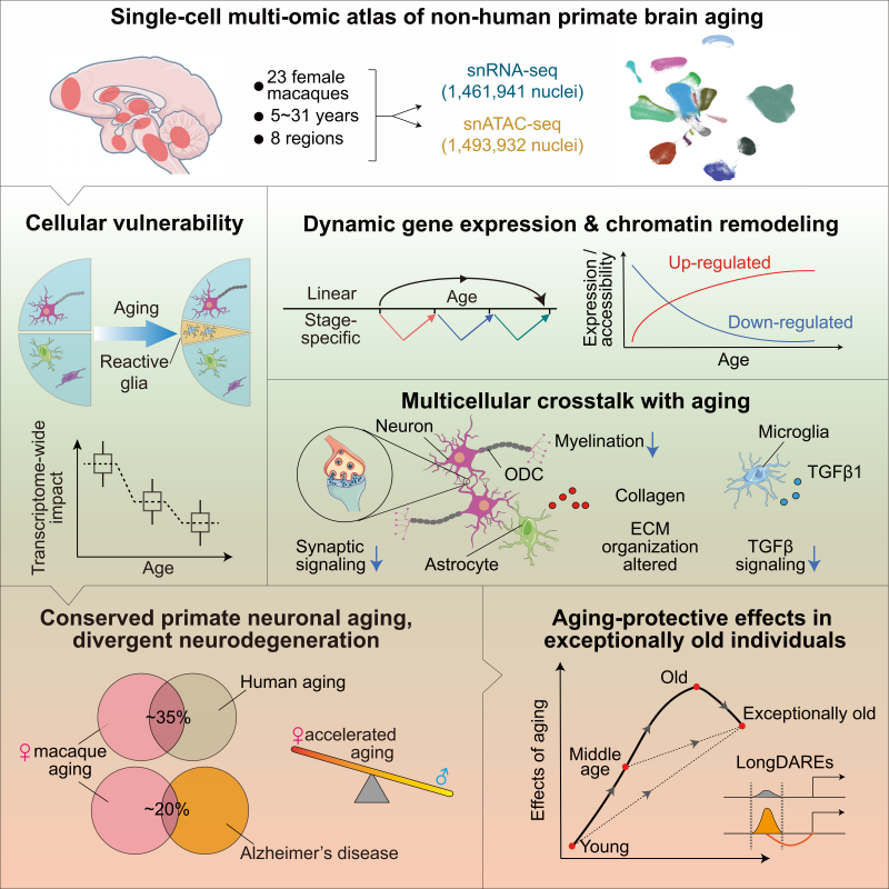 Eight brain regions from 23 female macaques aged 5 to 31 were profiled in two separate single-nucleus assays, giving a cell-by-cell reference for how the primate brain changes with age.
Research on primate brain aging has concentrated on the cortex, the outer sheet that does the brain’s higher-order work, and the cortex is not spared: prefrontal neuronal aging programs were the most coordinated of any region. But the heaviest burden of age-related change in gene expression turned up lower down, in the pons and medulla, a stretch of brainstem dense in white matter, the insulated long-distance wiring that relays traffic between the brain and the body.
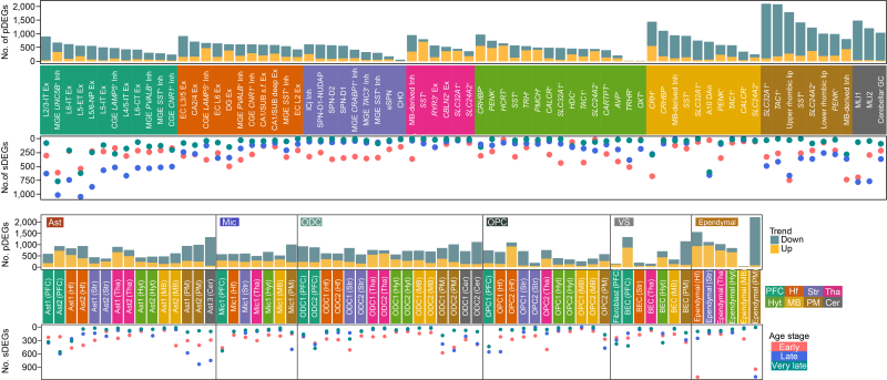 Number of age-related changed genes across cell subtypes, grouped by brain region, are highest in the pons and medulla, marking it as the focus of transcriptional change with age.
That burden was not carried by neurons alone. Recovered proportions of neurons stayed broadly stable with age, while glia, the support cells that feed neurons, police infection and maintain the myelin insulation around nerve fibers, changed sharply and by region, consistent with neuropathological evidence that age-related decline primarily reflects failing synapses, signaling and maintenance rather than widespread nerve-cell loss. In the pons and medulla those changes converged. Oligodendrocytes, which wrap nerve fibers in myelin much as insulation wraps a cable, showed their steepest losses of myelin genes, and the precursor cells meant to replace them pushed hardest here, a compensatory response that appears likely exhausted.
Knowing where the burden falls raises a second question: when does it arrive? Not evenly, and not mostly at the end. The most extensive transcriptional reorganization appeared in the comparison between young adult and middle-aged animals, then attenuated, and in the oldest group some changes had partly reversed. Because the four age groups are separate sets of animals, that shape is read across the cohort rather than within any one brain. It is not the smooth decline the word aging suggests.
The oldest animals were the surprise. Neurons in the exceptionally old macaques carried higher expression of genes for autophagy, the cell’s recycling system, plus protein folding and neurotransmitter release, the housekeeping functions that fall away elsewhere. Chromatin accessibility, which shows which pages of a cell’s instruction manual lie open, turned up regulatory elements enriched specifically in that group, including elements predicted to control BNIP3, a mitochondrial quality-control regulator linked to lifespan in fruit flies. The authors describe these as resilience-associated pathways that may help maintain neuronal function in late life, though the study cannot say whether that explains their longevity.
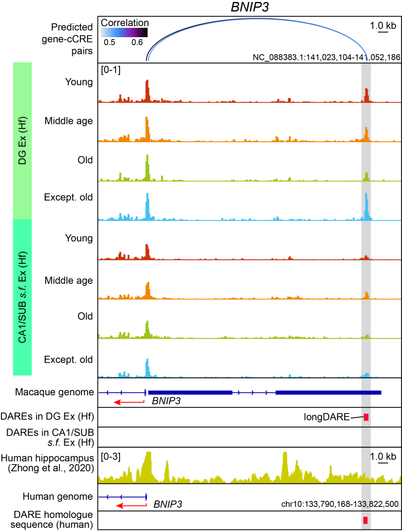 Chromatin accessibility at a regulatory element near BNIP3 is enriched in hippocampal neurons of the exceptionally old group, and the element has a conserved counterpart in the human genome.
Those elements belong to a larger map of close to 490,000 candidate regulatory elements, the switches that govern when nearby genes are used. Roughly nine in ten have homologous sequence in the human genome, and the smaller subset also conserved in human brain cells lets human genetic risk variants be tested against particular cell types. Variants associated with Alzheimer’s disease and multiple sclerosis were most strongly enriched in microglial regulatory elements, and those linked to white matter integrity in astrocytes and the oligodendrocyte lineage.
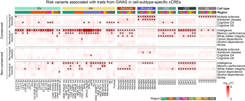 Human genetic risk variants for Alzheimer’s disease and multiple sclerosis are most enriched in microglial regulatory elements, and variants for white-matter integrity in astrocyte and oligodendrocyte-lineage elements.
Set against published human prefrontal cortex data, normal macaque aging in that region mirrors key transcriptional features of human aging, particularly in neurons. Alzheimer’s disease did not. It engaged partially distinct and sometimes opposite programs, which suggests that neurodegeneration is not simply normal aging on fast-forward.
In these animals the deepest reorganization came not at the end of life but on the way to middle age, and the oldest brains were not merely failing more slowly. What they were doing instead, and what sustains it, is the work the map now makes possible.
Ethics approval for this study is obtained.
The processed atlas is publicly available at a dedicated portal (https://db.cngb.org/stomics/nhpabc/), the raw data in CNGBdb-CNSA under accession number CNP0004459, and the code openly released (https://github.com/3DC-STAR-Anthony/NHPABC). This research is available at: https://doi.org/10.1016/j.cell.2026.07.045.